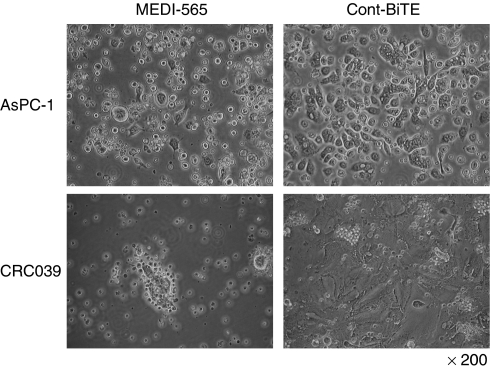
Figure 1.
MEDI-565 and T-cell (MEDI-565/T-cell)-mediated morphologic changes of CEA+ cancer cells. Carcinoembryonic antigen-positive human pancreatic cancer cell line, AsPC-1, and a CEA+ human colorectal cancer explant (CRC039) were used as target cells. T cells, negatively isolated from human PBMCs, were used as effector cells. 5 × 10⁵ target cells and 2.5 × 10⁶ T cells were co-cultured in each well of 12-well plates with 100 ng ml−1 of MEDI-565 or MEC14 control BiTE (Cont BiTE). Cells were incubated for 5 days at 37 °C, and photographed at × 200 magnification.

