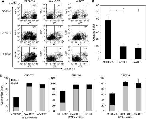
Figure 4.
MEDI-565/T-cell-mediated apoptosis of CEA+ human metastatic colorectal cancer explants. (A) Colorectal cancer cells from liver metastatic lesions, expanded NOD/SCID mice, excised and maintained in culture as described in the Materials and Methods, were incubated with MEDI-565 or MEC14 control BiTE (100 ng ml−1). After 5 days of incubation, cells were harvested with 0.05% trypsin/EDTA, washed and stained with biotin-annexin V and streptavidin-APC, FITC-anti-lineage marker, PE-anti-CEA and 7-AAD. Annexin V/7-AAD positivity was evaluated in lineage marker-negative, CEA+ tumour cell populations using flow cytometry. The percentages of annexin V-positive cells are shown in each dot plot. (B) Co-incubation of T cells and colorectal cancer cells (CRC007, CRC010 and CRC039) together with MEDI-565 or Cont BiTE was repeated twice, and the average percentages of apoptotic tumour cells (Annexin V+) are shown in the bar graph. *P<0.005 (Student's t-test). (C) Cells were counted with Trypan blue dye exclusion method. The numbers of viable cells (grey) and dead cells (black) are shown in each bar graph. The error bars show s.d. of the total number of cells calculated from duplicate wells.

