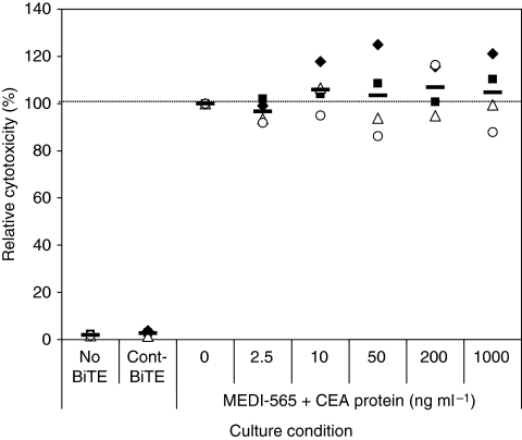
Figure 5.
Soluble CEA protein does not affect MEDI-565/T-cell-mediated apoptosis of CEA+ cell line and colorectal cancer cells. AsPC-1 cells or CRC010 colorectal cancer cells (5 × 10⁵ cells per well) were incubated with T cells (2.5 × 10⁶ cells per well) in a 12-well plate in the presence of MEDI-565 (100 ng ml−1; 1.8 nM) with the indicated concentration of soluble CEA antigen in the medium (2.5∼1000 ng ml−1; 0.01 to 5 nM). After 5 days of incubation, tumour cells were harvested and stained as described in the Figure 4 legend. Annexin V-positive cells in lineage marker-negative, CEA+ tumour cells were evaluated. The cytotoxicity value of the culture with MEDI-565 without soluble CEA protein was set as 100%, and each cytotoxicity value is shown as a percentage relative to this MEDI-565-positive, CEA protein-negative culture. The assay was repeated twice for both AsPC-1 cells and CRC010 cancer cells (filled symbols indicate AsPC-1 cells and open symbols indicate CRC010 tumour cells; each symbol represents an individual T-cell donor). The bars show the average cytotoxicities of each condition.

