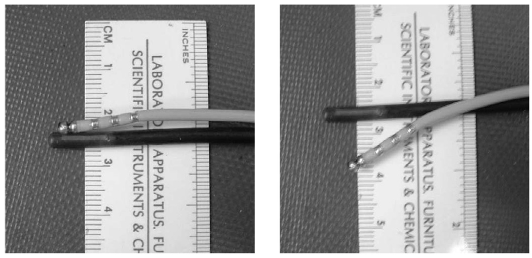
Fig. 1.
Coupled imaging and ablation (with metallic rings) catheters used at the second and third ablation sites. Joining the two probes facilitated catheter position and alignment within the heart. The two probes are fixed in their “parallel” position (a) when traveling through vessels. When properly positioned with the heart, the ablation catheter can be flexed downward (b) and onto the target myocardium for an ablation.

