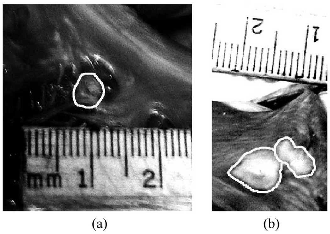
Fig. 9.
Images from pathology of the inner myocardial surface of the right atrium containing radiofrequency-generated lesions. The lesion boundaries were manually outlined. The first lesion (a) was located within the right atrial appendage. The second and third lesions were side-by-side lesions found on the right atrial free wall near the opening to the superior vena cava.

