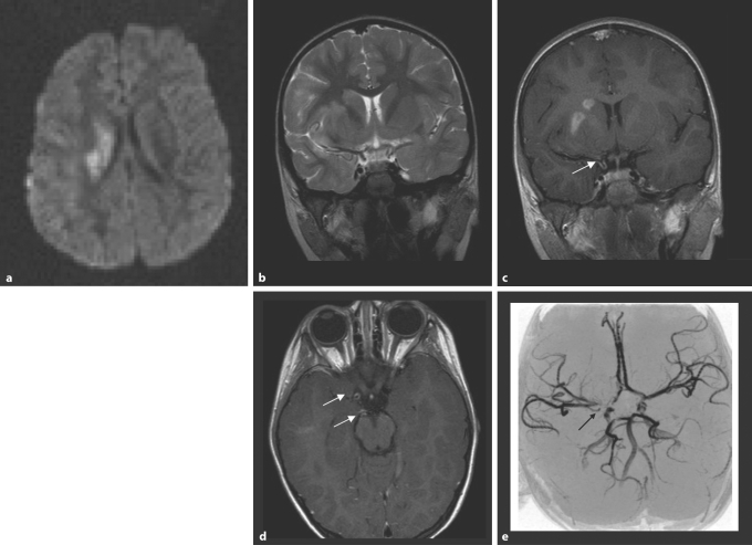
Fig. 1.
cPACNS; mild right hemispheric stroke with hemiparesis 10 days earlier; 4-year-old girl. a DWI. There is restricted diffusion of protons in the right basal ganglia. b The T2-weighted MRI shows only minor hyperintense signal change in the basal ganglia on the right sparing the internal capsule. c The contrast-enhanced coronal T1-weighted image (slice thickness 3 mm, flow compensation) after contrast injection (0.1 mmol/kg Gd-DTPA) through the distal internal carotid artery shows contrast enhancement in the right caudate and lentiforme nuclei. There is clearly enhancement in the wall of the right distal internal carotid artery (arrow). d The axial T1-weighted image shows wall enhancement in the right distal internal carotid artery and the right P1 segment (arrow). e The TOF-MRA shows an abnormality of the right internal carotid artery around the carotid T, but also some narrowing in the right P1 (arrow).

