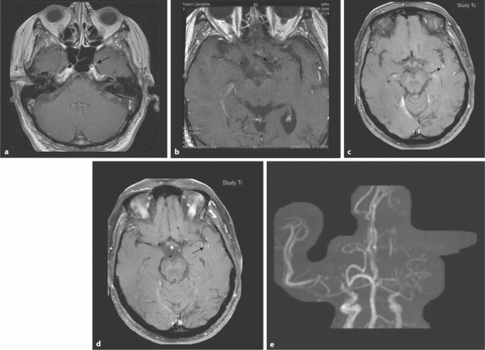
Fig. 2.
Fifty-three-year-old woman with right-sided hemiparesis and aphasia. a–d High-resolution T1-weighted images after contrast injection (0.1 mmol/kg) with fat suppression and flow compensation. a This T1-weighted contrast-enhanced image at the level of the skull base shows an enlargement of the left distal internal carotid artery (arrow) compared to the right. b At a slightly higher location, there is contrast enhancement in the M1 segment on the left (arrow). c Wall enhancement is also seen in middle cerebral artery branches in the left sylvian fissure (arrow). d This image slightly more apical confirms the intramural enhancement pattern (arrow). e The TOF-MRA shows severe flow abnormality in the left distal internal carotid artery and A1 as well as M1 segments. There are also severe stenoses on the right.

