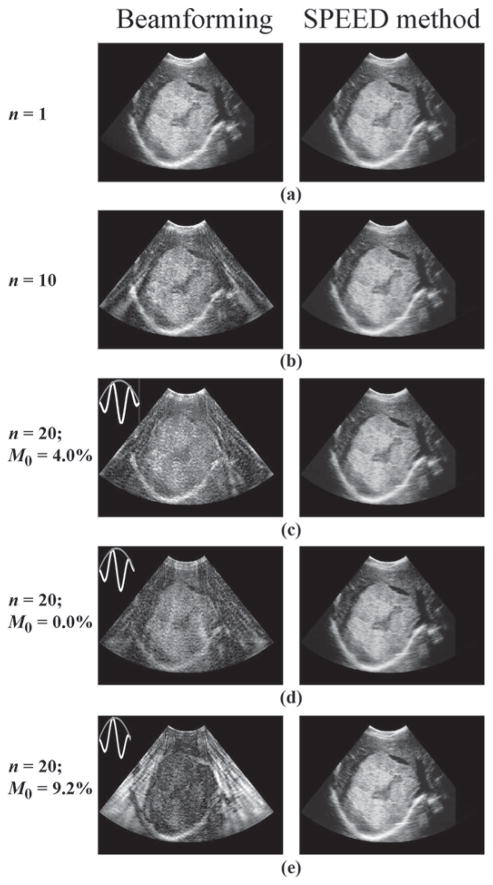
Fig. 7.
A clinical liver image showing a large hemangioma with cystic degeneration was sampled along 120 different beams to translate the object to polar coordinates. The raw RF data were then simulated through (1), and data from different beams were combined to simulate acceleration factors of 1, 10, and 20. The resulting data sets were reconstructed using receive beamforming and the proposed spatial-encoding approach of (2), (3), and (5). Although accelerated results obtained with a beam-forming reconstruction featured artifacts, results from the proposed approach were essentially artifact-free. The quality of beamforming results was found to depend greatly on the waveform used for the transmitted waveform, and the n = 20 results were repeated for waveforms featuring slightly different envelopes. A normalized version of the zeroth moment, M0 = |∫w(t)dt|/∫|w(t)|dt with w(t) the transmitted waveform, was used to characterize the waveforms. The quality of images obtained with a receive beamforming reconstruction tended to decrease with increasing values of M0. A main purpose of this simulation was to demonstrate that neither object complexity nor M0 adversely affects the proposed algorithm.

