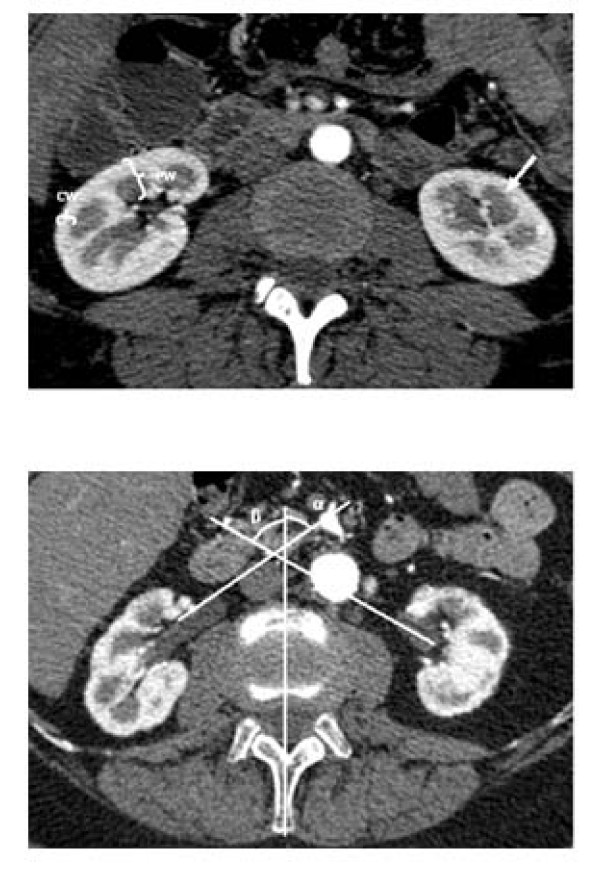
Figure 1.
Axial 0.625 mm collimated slice of the kidney in an arterial phase, with the strongly contrasted kidney cortex (*), and a renal pyramid (arrow). Cortical width (CW), and parenchymal width (PW) (a). Axial 0.625 mm collimated slice of the kidney in an arterial phase, with depiction of the kidney pelvis, and the rotation status of the kidney pelves in relation to the reference sagittal median plane (b). Pelvic angle on the right side (α), and on the left side (β).

