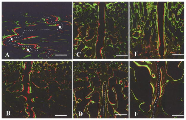
Fig. 5.
Mineralization at suture margins and suture width. A) Interfrontal and B) interparietal suture of a 3-month-old pig (#297); C) interfrontal and D) interparietal suture of a 7-month-old pig (#306). Suture mineral apposition rate (MAR) was greater in younger animals. E) Ectocranial and F) endocranial regions of the unfused interparietal suture of a 7-month-old pig (#322). The average width was greater on the ectocranial side. Arrows in (A) indicate high growth rate at the tips of interdigitations. For clarity, the suture space in (A), (D) and (F) are marked by broken lines. To scale: calibration bar, 500 μm.

