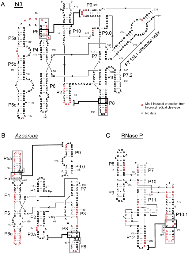
Figure 3. Secondary structure models illustrating Mrs1 protections for the bI3, Azoarcus, and RNase P RNAs.
Nucleotides protected from hydroxyl radical cleavage upon Mrs1 binding are red. Bold black lines emphasize tetraloop-receptor interactions; protected residues adjacent to the receptor helices are emphasized with gray boxes. Paired helical regions are indicated by Px. Group I intron splice sites are indicated with open arrows. A small number of sites with no data, due to high background, are shown in gray lettering.

