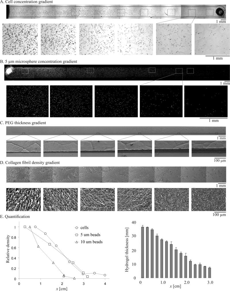
Figure 3. Long-range gradients of particles, cells and materials.
SEM images at low and high magnification of freeze-dried A) PEG-DA hydrogel gradient and B) collagen gradient. Microscope images of C) endothelial cell gradient and D) fluorescent particle gradient (diameter 5 μm). E) Quantification of the continuous variance in thickness of the hydrogel gradient and relative density profiles of endothelial cell gradient and fluorescent particle gradient (with diameters of 5, 10 μm).

