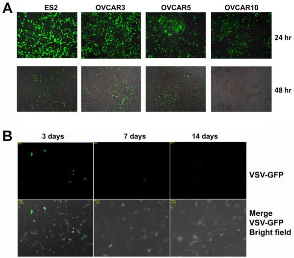
Fig. 2.
Oncolytic activity of VSV-GFP in cultured ovarian epithelial and tumor cells. (A) Ovarian cancer cells ES2, NIH:OVCAR5, and A2780 in 24-well dishes were infected with 106 pfu of VSV-GFP. The cells were observed for GFP expression and for PI staining (red) to mark dead cells 2 days after infection. (B) Primary ovarian surface epithelial cell preparation HOSE-65 cells in 24-well dishes were infected with 108 pfu of VSV-GFP and monitored for GFP expression on day 3, 7, and 14 after infection.

