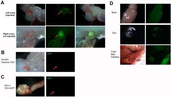
Fig. 3.
Targeting of VSV following intra ovarian bursa injection in Wv mice. Four-month-old Wv/Wv mice that bear ovarian tumors were injected with 105 pfu of VSV-GFP into the bursa of left ovary. Ten days after injection, the mice were dissected and organs were examined under fluorescence microscopy for GFP signals. (A) The injected left ovary of a representative tumor-bearing Wv mouse shows fluorescence (arrow). The non-injected right ovary from the same mouse also shows GFP fluorescence. As controls, no signals were detected in ovaries in Wv/Wv mice injected with inactivated VSV (B), or in Wv heterozygous (no ovarian tumor) mutant mice (C). (D) Other organs (brain, eye, and liver/bile/intestine are shown) from VSV-GFP-injected mice show no significant GFP signals.

