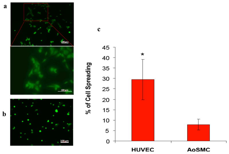
Figure 5.
Fluorescent images of (a) HUVECs and (b) AoSMCs on PA-YK after 2 hours using Live/Dead assay. HUVECs attain their regular spread morphology within 2 hours. AoSMCs do not display any signs of spreading. (c) Initial spreading of HUVECs and AoSMCs on PA-YK nanofibrous matrix. HUVECs show significantly greater spreading than AoSMCs after 2 hrs (*p<0.05). Error bar represents means ± standard deviation for n=12.

