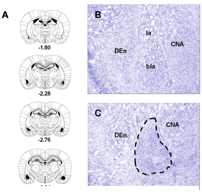
Fig. 1.
Series of reconstructions (A) of the largest (gray) and the smallest (black) lesions of the basolateral amygdala (BLA) complex at four coronal levels (−1.80, −2.28, −2.76, −3.24 mm posterior to bregma; the diagrams were adapted with permission from plates in Paxinos and Watson [38] atlas). Digital photomicrographs of coronal brain sections at the level of the amygdala of a neurologically intact subject (B) and a rat with excitotoxic lesions of the BLA (C; the dashed lines shows the extent of cell loss). Abbreviations: bla, basolateral amygdaloid nucleus; CNA, central nucleus of the amygdala; DEn, dorsal endopiriform nucleus; la, lateral amygdaloid nucleus.

