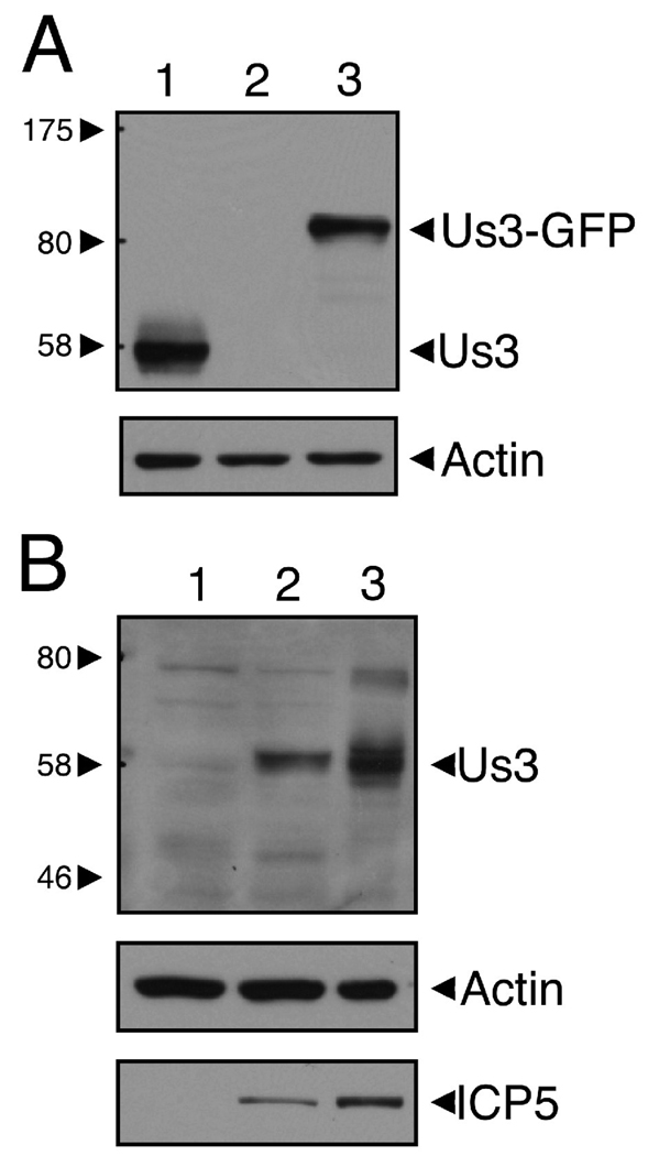
Figure 1.
Polyclonal antiserum from rats immunized with GST-HSV-2 Us3 specifically detects HSV-2 Us3. A. Equal volumes of cellular extracts prepared from 293T cells transfected with a plasmid encoding HSV-2 Us3 (lane 1), GFP (lane 2), or HSV-2 Us3-GFP (lane 3) were analyzed by Western blotting. The upper panel was probed with rat polyclonal HSV-2 Us3 antiserum and the loading control in the lower panel was probed with anti-actin monoclonal antibody. No bands were detected in lane 2 of the upper panel (including bands smaller than 58 kDa, which are not shown). The major band detected in lane 1 is slightly larger than the predicted molecular weight of HSV-2 Us3 (53 kDa). Note the shift in molecular weight of the detected band in cells transfected with plasmid encoding HSV-2 Us3-GFP fusion protein. B. Equal volumes of cellular extracts prepared from mock-infected (lane 1), HSV-1 17+-infected (lane 2), or HSV-2 HG52-infected Vero cells (lane 3) were analyzed by Western blotting. The upper panel was probed with rat polyclonal HSV-2 Us3 antiserum, the loading control in the middle panel was probed with anti-actin monoclonal antibody, and the infection control in the lower panel was probed with anti-HSV-2 ICP5. Molecular size markers indicated at the left of each upper panel are in kilodaltons.

