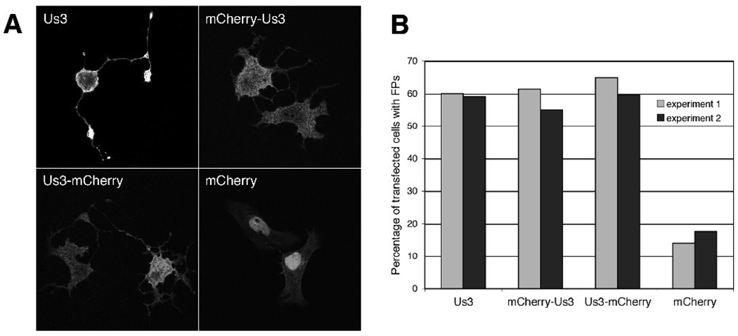
Figure 2.
Expression of HSV-2 Us3 in transfected cells results in FP formation. A. Representative images of FPs formed in Vero cells transfected with plasmids encoding HSV-2 Us3, mCherry fusions to HSV-2 Us3 or mCherry alone. In the upper left panel, staining with polyclonal antiserum specific for HSV-2 Us3 followed by staining with an Alexa 488 conjugated secondary antibody was used to detect cells expressing HSV-2 Us3. Stained cells were examined by confocal microscopy. B. Percentage of transfected cells with FPs. 50 independent fields of cells containing at least one transfected cell were scored for the presence or absence of FPs. Results from two independent experiments are shown. Numbers of cells scored in experiment 1 and experiment 2 were as follows: for WT Us3 – 115 and 127; for mCherry-Us3 – 145 and 156; for Us3 mCherry – 168 and 154; for mCherry – 206 and 164.

