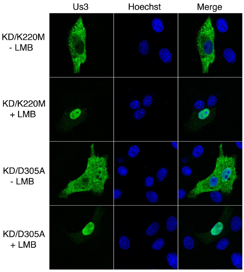
Figure 9.
Subcellular localization of HSV-2 Us3 in the presence of LMB. Vero cells were transfected with plasmids encoding the indicated proteins. At 6 hours post transfection cells were placed in medium containing 10 ηM LMB (+ LMB) or 0.07% methanol carrier (− LMB). Cells were incubated in the continuous presence of drug or carrier for 16 hours, then fixed and stained with rat polyclonal antiserum specific for HSV-2 Us3 followed by staining with an Alexa 488 conjugated secondary antibody. Nuclei were stained with Hoechst 33342. Stained cells were examined by confocal microscopy. A large proportion of transfected cells treated with LMB showed the staining pattern depicted in the “+ LMB” panels.

