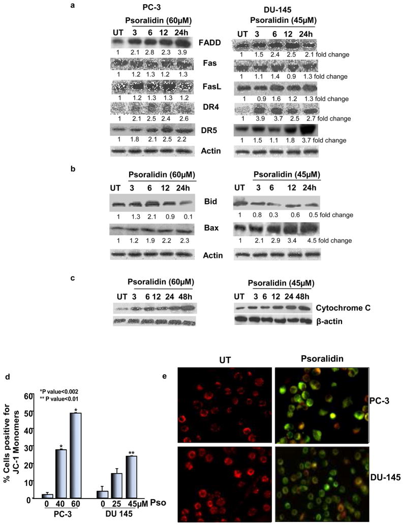
Figure 3. Effect of psoralidin on death receptor and apoptotic signaling in AIPC cells.
PC-3 and DU-145 cells (70–80% confluency) were treated with 60 and 45 μM psoralidin, respectively, for varying time intervals (3–24 h), and Western blot analysis was performed using (A) FADD, Fas, FasL, DR4 and DR5 antibodies and (B) Bid and Bax antibodies. Actin was used as the internal loading control. (C) PC-3 and DU-145 cells (70–80% confluency) were treated with 60 and 45 μM psoralidin, respectively, for varying time intervals (3–24 h), and the cytoplasmic fraction of control and psoralidin treated cells were subjected to Western blot analysis using a cytochrome C antibody. (D) PC-3 and DU-145 cells (70–80% confluency) were treated with 40 or 60 μM and 25 or 45 μM psoralidin respectively for 24 h and JC-1 staining was performed using flowcytometry. Bars represent percentage of cells positive for green fluorescence (JC-1 monomers) ±SD. (E) PC-3 and DU-145 cells plated in 8-well chamber slides (70–80% confluency) were treated with 60 and 45 μM psoralidin, respectively, for 24 h, and JC-1 staining was performed and slides were analyzed by confocal microscopy.

