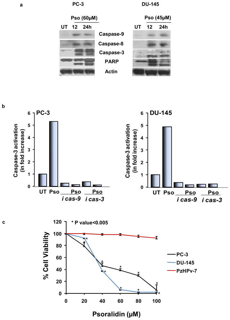
Figure 4. Effect of psoralidin on caspase signaling and viability of AIPC cells.
(A). PC-3 and DU-145 cells (70–80% confluency) were treated with 60 and 45 μM psoralidin, respectively, for 12 and 24 h, and Western blot analysis was performed using caspase-9, caspase-8, caspase -3 and PARP antibodies. Actin was used as the internal loading control. (B). PC-3 and DU-145 cells (70–80% confluency) were treated with psoralidin alone, caspase inhibitors alone (3 or 9) and a combination of caspase inhibitors and psoralidin, and a fluorometric assay was performed to determine caspase-3 activation. Bars represent fold increase in caspase activity in each treatment group. (C). PC-3, DU-145 and PzHPv-7 (70–80% confluency) cells were treated with varying concentrations of psoralidin, and cell viability was determined using Trypan blue assay. A dose-response curve was plotted, and each data point represents mean percentage of cell viability±SD.

