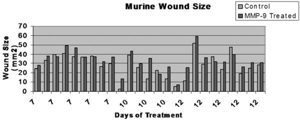
Fig. 3. Wounds.
25 Mice were sacrificed at three time points, 7 10 and 12 days. The dorsal skin and underlying muscle were excised, placed on a flat sheet of paper and photographed from a set distance. Here control wounds are picture in blue and MMP-9 exposed wounds in red, paired by animal. Inter-animal variability is notable, but MMP-9 exposed wounds are consistently larger then their paired treatment wound.

