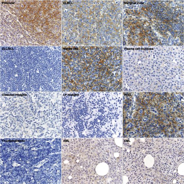
Figure 3. Immunohistologic staining for CD81 in hematolymphoid neoplasia.
Representative examples of CD81 immunostaining in lymphomas (60x magnification) show CD81 expression in follicular lymphoma, diffuse large B cell lymphoma, marginal zone lymphoma, mantle cell lymphoma, and peripheral T cell lymphoma. CD81 staining was absent in small lymphocytic lymphoma/chronic lymphocytic leukemia, plasma cell myeloma, Hodgkin lymphoma, T-lymphoblastic lymphoma, acute myeloid leukemia, and chronic myeloid leukemia.

