
Figure 2.
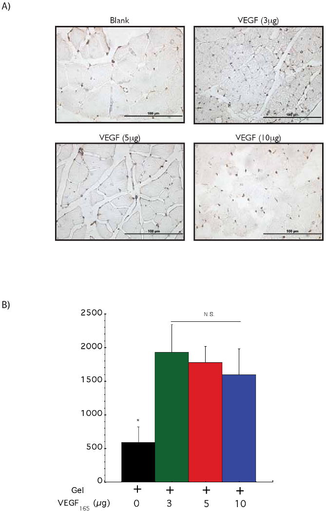

Monitoring the effect of VEGF dose in driving vascularization in ApoE-/- ischemic hindlimbs. Representative photomicrographs from CD31 immunostained sections of hindlimb muscle tissues (A). Quantification of blood vessel densities in hindlimb muscle tissues 6 weeks after treatment with a control blank gel (+ 0); a gel with 3 μg of VEGF (+ 3); a gel with 5 μg of VEGF (+ 5) and a gel with 10 μg of VEGF (+ 10) (B). No statistically significant differences were observed between the different VEGF doses. Tissue perfusion of ApoE-/- mice hindlimbs at various time points following treatment with a control blank gel (◇); 3 μg of VEGF delivered from alginate hydrogels (●); 5 μg of VEGF delivered from alginate hydrogels (○) and 10 μg of VEGF delivered from alginate hydrogels (□) (C). Mean values are presented with standard deviations and * indicates statistically significant differences (p<0.05), as compared to control gels. N.S. displays no statistically significant difference between conditions.