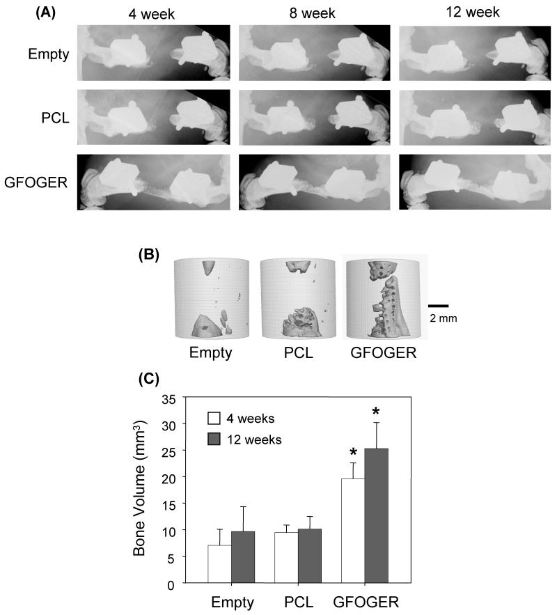
Figure 3.
GFOGER-coated scaffolds significantly enhance bone formation in critically-sized defects compared to uncoated scaffolds and empty defect controls. (A) Faxitron images show that empty defects do not heal after 12 weeks, and negligible bone formation is present in uncoated PCL-treated defects. However, GFOGER-treated defects promote bone formation as early as 4 weeks. (B) MicroCT images for samples shown in (A) at 12 weeks. (C) Bone volume is significantly greater in GFOGER-treated samples at both 4 and 12 weeks compared to empty defects and uncoated PCL-treated samples. Error bars represent standard error of the mean (n=8 and n=9 for PCL and GFOGER at 4 weeks, respectively; n=7 and n=8 for PCL and GFOGER at 12 weeks, respectively). * Different from empty defect and PCL (p<0.05).

