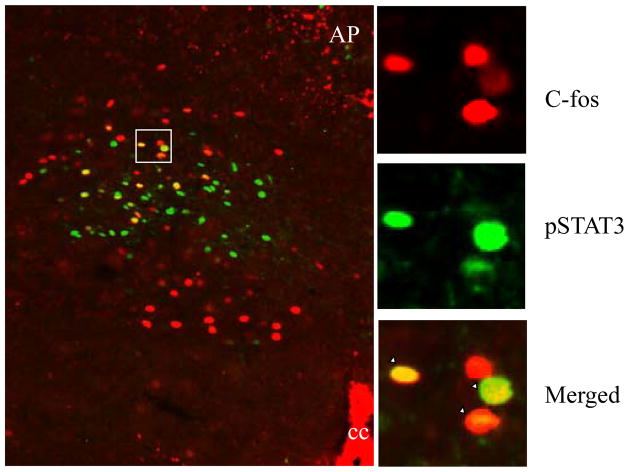
Figure 2.
A significant proportion of leptin-responsive cells respond to gastric distension. A representative merged microphotograph of double IHC (P-STAT3 and c-Fos) from gastric distension combined with leptin-treated rats is shown on the left. On the right are shown high magnifications (top, P-STAT3 green fluorescence IHC; middle, c-Fos red fluorescence IHC; bottom, merged microphotograph from the double IHC) of the area marked on the left. Examples of double-labeled cells are shown in yellow. cc, Central canal. Scale bars, 200 μm.

