Figure 1.
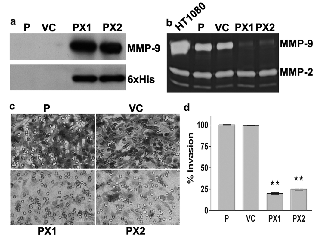
Inhibition of MMP-9 activity and tumor cell invasion by MMP-9 PEX. (a) Western Blotting. SNB19 cells were transfected with either vector alone (VC) or MMP-9-PEX vector and stable transfectants (PX1, PX2) were selected. Conditioned media were analyzed by western blotting using anti-MMP-9 antibody or anti-His tag antibody. (b) Gelatin zymography. Parental and stable transfectants were cultured in growth medium supplemented with 200 nM phorbol myristate acetate (PMA) for 24-h. Conditioned medium obtained from PMA-treated parental (P), vector alone (VC) and MMP-9 PEX stable transfectants (PX1, PX2) and were resolved under nonreducing conditions on 10% SDS-PAGE gels. Gels were stained with amido black and areas of gelatinolysis were visualized as transparent bands. Conditioned medium from HT1080 cells was used to identify the gelatinolytic bands and their approximate molecular weights on the gels. (c) Matrigel invasion assay. Transfected SNB19 cells were assayed for invasive capacity in Matrigel coated tissue culture inserts for 24-h. Cells migrated to the bottom side of the insert were stained and photographed. Representative images are shown from one of three independent experiments with similar results. (d) Invasion index was determined as mean ± SD of three experiments done in duplicate. **p< 0.01 significantly different from vector alone transfected cells.

