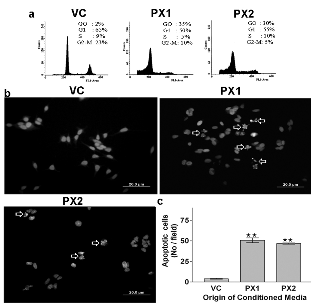
Figure 5.
Flow cytometry. (a) Effect of MMP-9-PEX on cell cycle distribution in HMECs. Cells were grown in vector alone (VC) or MMP-9-PEX SNB19 (PX1, PX2) cell conditioned medium for 24 hr and then the cells were harvested, stained with propidium iodide and cell cycle distribution was determined by flow cytometry. A representative cell cycle analysis is shown. Apoptosis evaluation. (b) Apoptosis was evaluated in HMECs at 24-h after exposure to vector alone (VC) or MMP-9-PEX SNB19 (PX1, PX2) cell conditioned medium by fluorescence microscopy using the chromatin stain Hoechst 33258. (c) The apoptotic index was quantitated by counting five random fields (in duplicate wells) per group. Data represent a result from two independent experiments. **p< 0.01

