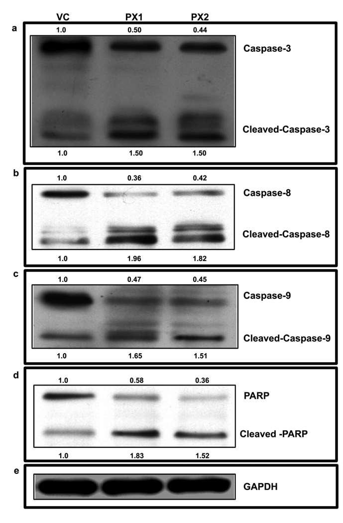
Figure 6.
Effects of PEX on apoptosis in HMECs. Endothelial cells were grown in vector alone (VC) or MMP-9-PEX SNB19 (PX1, PX2) cell conditioned medium for 24-h harvested and cell lysates were subjected to electrophoretic analysis through SDS-PAGE followed by transfer to nitrocellulose membrane. Western blot analysis was done with the indicated antibodies and representative samples were shown.

