Figure 7.
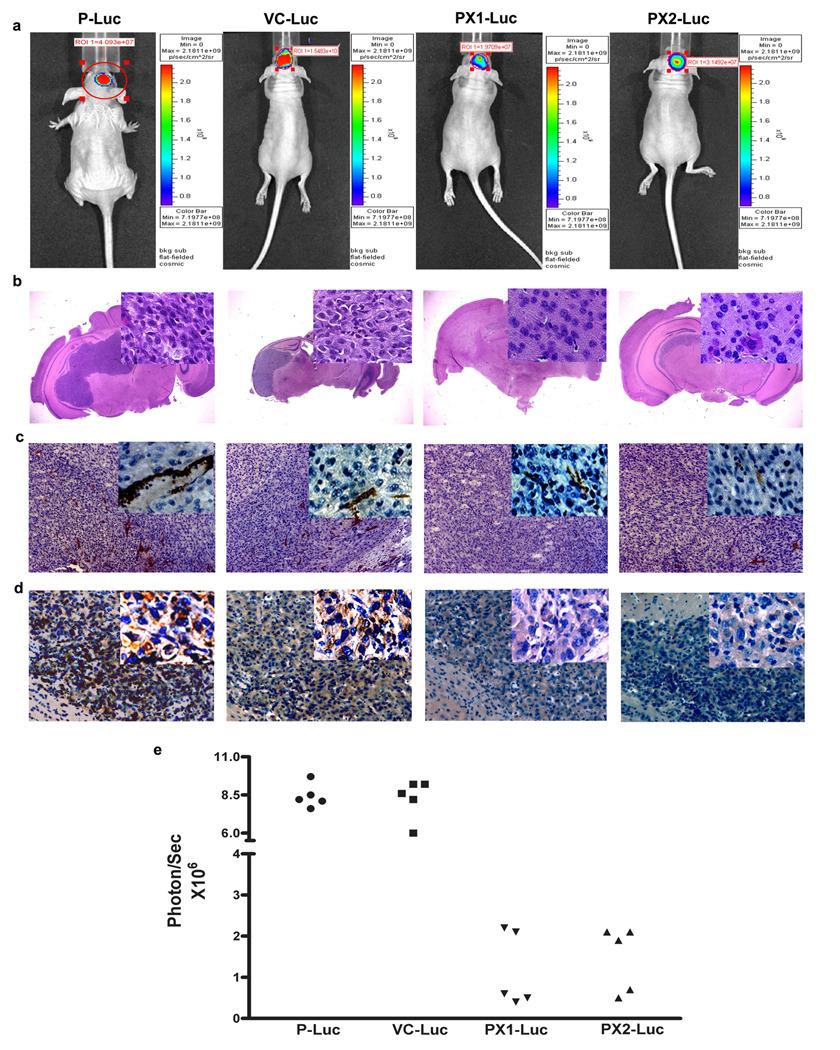
Effect of MMP-9-PEX on the growth of intracranial SNB19 tumors (a) Bioluminescence imaging of SNB19 tumors in vivo. Parental (P), vector alone (VC) and MMP-9-PEX SNB19 transfectants (PX1, PX2) engineered to express luciferase by phCMV-Luc transfection were examined by optical imaging. A representative experiment shows the week four images of bioluminescence being obtained simultaneously from intracranial tumors of mice implanted of parental (P-Luc), vector alone (VC-Luc) or MMP-9-PEX SNB19 cells (PX1-Luc, PX2-Luc). (b) Photomicrograph of H&E staining from tumor sections. Insert, area that was magnified. (c) Immunohistochemistry of CD31 on xenografted tumors from parental SNB19 cells and stable transfectants (d) Analysis of xenografted tumors by immunohistochemical staining using anti-MMP-9 antibody (e) Bioluminescence data corresponding to individual mice from animals intracranially implanted with parental (P-Luc, n = 5), vector alone (VC-Luc, n = 5) and MMP-9-PEX vector transfected (PX1-Luc, n = 5; PX2-Luc, n = 5) glioblastoma cells.

