Figure 2.

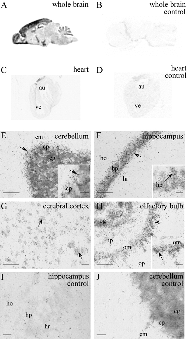
PIP5K2C gene expression in adult mouse tissues by in situ hybridization. With PIP5K2C-specific probes on 20-μm mouse tissue sections, positive signal is spatially distributed in brain (A) but absent from heart tissue (C; not to scale). Control sections of brain (B) and heart (D) were incubated with excess of unlabeled probe, and identical results were obtained with three sections from two animals. Autoradiographic emulsion staining of hybridization slides (counterstained with methyl blue), shows PIP5K2C mRNA labeled with silver grains (E–J). Positive cells (arrows) are seen in the cerebellum (E, inset is Purkinje cell layer), hippocampal field CA1 (F, inset is stratum pyramidale), cerebral cortex (G, inset is layer V), and olfactory bulb (H, inset is mitral cell layer). Controls incubated with excess cold probe (I,J) were negative. au, Heart auricle; ve, heart ventricle; cm, molecular layer; cp, Purkinje layer; cg, granular layer (all cerebellar); ho, stratum oriens; hp, stratum pyramidale; hr, stratum radiatum (all hippocampal); op, outer plexiform layer; om, mitral cell layer; ip, inner plexiform layer; og, granule cell layer (all olfactory bulb). Scale bars = 40 μm in E–I; 10 μm in J and insets.
