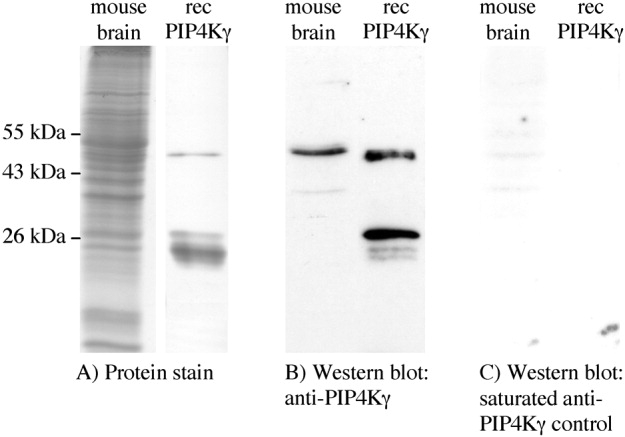
Figure 3.

Specificity of antiphosphatidylinositol 5-phosphate 4-kinase γ (PIP4Kγ) antibody to PIP4Kγ in mouse brain. A: SDS-PAGE of total proteins from 50 μg whole mouse brain lysate (Coomassie stained) and 50 ng of purified recombinant (rec) PIP4Kγ (silver stained) showing mature protein of 47 kDa and degradation products at 26 kDa. B: Western blot of these proteins using anti-PIP4Kγ antibody, showing single band specificity in brain lysate. C: Control Western blot using anti-PIP4Kγ antibody preincubated with antigenic peptide. Results are representative of at least three experiments.
