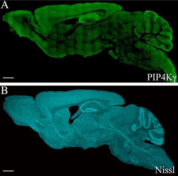
Figure 6.

PIP4Kγ expression in different regions of the adult mouse brain. PIP4Kγ was detected in whole saggital brain sections (approximately lateral 0.48 mm) by immunofluorescence with anti-PIP4Kγ antibody (A). Staining indicated differential expression levels of PIP4Kγ in different brain regions. A similar, fluorescent Nissl-stained section is included for morphological reference (B). Images are representative of a minimum of six stained sections from three different mice. Scale bars = 1 mm. [Color figure can be viewed in the online issue, which is available at www.interscience.wiley.com.]
