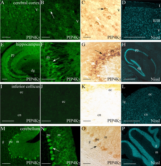
Figure 7.
A–P: Localization of PIP4Kγ in adult mouse brain regions. Positive PIP4Kγ signal (arrows) was observed in the cerebral cortex (A–C), CA1–CA3 of the hippocampus (E–G) and cerebellum (M–O). No significant signal was observed in the inferior colliculus (I–K). Saggital brain sections were stained by immunofluorescent (A,B,E,F,I,J,M,N) and also immunochemical (C,G,K,O) methods. Fluorescent Nissl-stained images are included for reference (D,H,L,P). Images are representative of a minimum of six stained sections from three different mice. I, II/III, V, cerebral cortex layers; dg, dentate gyrus; py, hippocampal pyramidal cells; ec, external cortex of the inferior collicus; cn, central nucleus of the inferior collicus; ig, intermediate gray layer of the superior collicus; pu, cerebellar Purkinje cells; g, cerebellar granular layer; m, cerebellar molecular layer. Scale bars = 100 μm in A,E,I,M; 50 μm in B,F,J,N; 20 μm in C,G,K,O; 500 μm in D,H,L,P. [Color figure can be viewed in the online issue, which is available at www.interscience.wiley.com.]

