Figure 8.
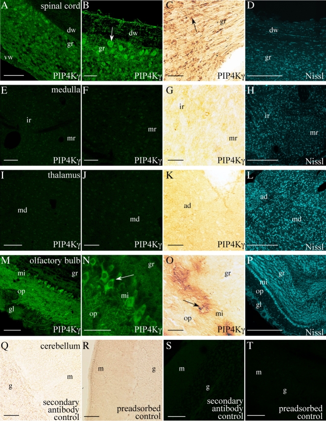
A–T: Localization of PIP4Kγ in adult mouse brain regions. Positive PIP4Kγ signal (arrows) was observed in the cervical spinal cord (longitudinal section, A–C) and olfactory bulb (M–O). No significant signal was observed in other areas such as the medulla (E–G) or thalamus (I–K). Saggital brain sections were stained by immunofluorescent (A,B,E,F,I,J,M,N,S,T) and also immunochemical (C,G,K,O,Q,R) methods. Fluorescent Nissl-stained images are included for reference (D,H,L,P). Controls for immunochemical and immunofluorescent labeling (incubated with secondary antibody only or using preadsorbed anti-PIP4Kγ) are also included (Q–T). Images are representative of a minimum of six stained sections from three different mice. dw, Dorsal white matter of the spine; vw, ventral white matter of the spine; gr, spinal gray matter; ir, intermediate reticular nucleus of the medulla; mr, medullary reticular nucleus; md, mediodorsal/paracentral thalamic nuclei; ad, anterodorsal thalamic nucleus; gl, olfactory glomerular layer; op, olfactory outer plexiform layer; gr, olfactory granule cell layer; mi, olfactory mitral cells; g, cerebellar granular layer; m, cerebellar molecular layer. Scale bars = 100 μm in A,E,I,M; 50 μm in B,F,J,N; 20 μm in C,G,K,O,Q–T; 500 μm in D,H,L,P. [Color figure can be viewed in the online issue, which is available at www.interscience.wiley.com.]

