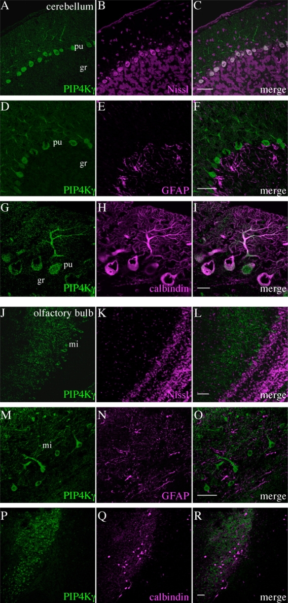
Figure 10.

Identification of cell types expressing PIP4Kγ in the adult mouse cerebellum and olfactory bulb. PIP4Kγ (green) was detected in the cerebellum (A–I) and olfactory bulb (J–R). Signal was coincident with fluorescent Nissl stain (A–C,J–L) and was excluded from glial cells stained with anti-GFAP (D–F,M–O), indicating that PIP4Kγ was expressed in neuronal cells. PIP4Kγ was expressed in cells that were positive for the neuronal cell marker calbindin D-28k in the cerebellum (G–I) but not in the olfactory bulb (P–R). Images are representative of three stained sections. gr, Cerebellar granular layer; pu, cerebellar Purkinje layer; mi, mitral cell layer of olfactory bulb. Scale bars = 50 μm in C (applies to A–C); 50 μm in F (applies to D–F); 20 μm in I (applies to G–I); 50 μm in L (applies to J–L); 20 μm in O (applies to M–O); 50 μm in R (applies to P–R).
