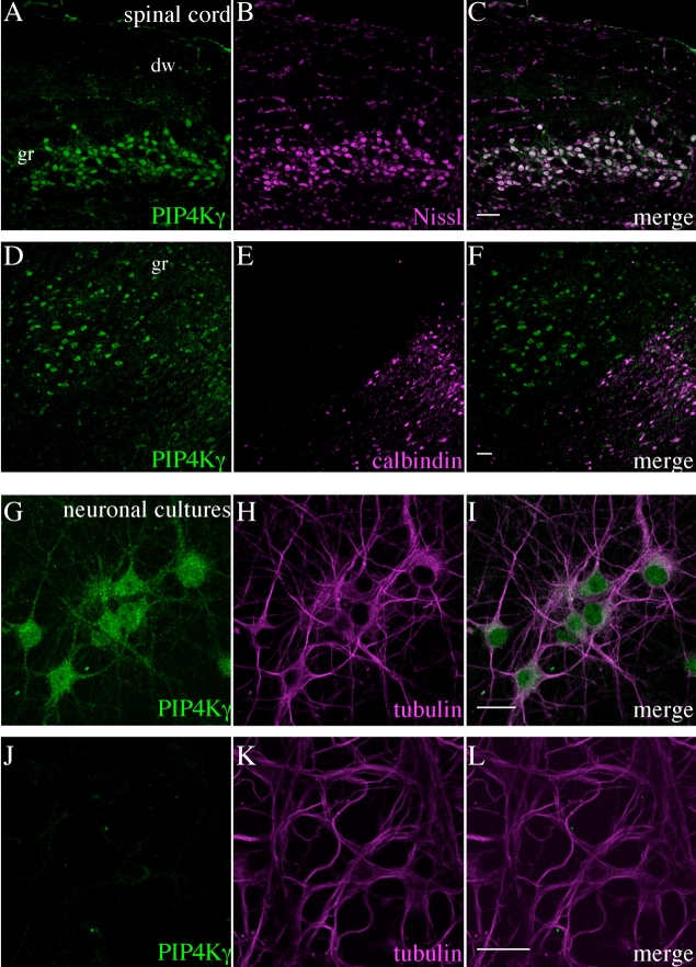
Figure 11.
Identification of cell types expressing PIP4Kγ in the adult mouse spinal cord and in primary neuronal cultures. Endogenous PIP4Kγ (green) was detected in neurons in the spinal cord (A–F) by fluorescent costaining for Nissl substance (A–C) and was also seen in the subpopulation of neurons positive for calbindin D-28k (D–F). Hippocampal primary cell cultures enriched for pyramidal cells expressed PIP4Kγ (G–I), whereas cultures enriched for cerebellar granule cells did not (J–L). Images for (A–F) are representative of three stained sections. dw, Dorsal white matter of the spine; gr, spinal gray matter. Scale bars = 50 μm in C (applies to A–C); 50 μm in F (applies to D–F); 20 μm in I (applies to G–I); 20 μm in L (applies to J–L).

