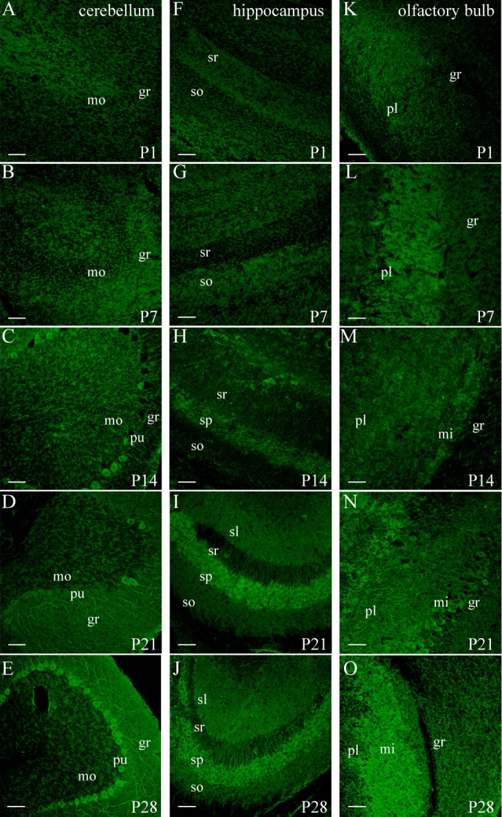
Figure 14.

Expression of PIP4Kγ in the developing mouse brain. Immunohistochemical staining of mouse brain sections at different postnatal development stages (P1–P28 days after birth) indicated the levels of PIP4Kγ (green) detected in three different regions; cerebellum (A–E), hippocampal field CA3 (F–J), and olfactory bulb (K–O). Brain regions are pictured in the same orientation, and images are representative of two animals at each developmental stage. mo, Molecular layer; pu, Purkinje layer; gr, granular layer (cerebellum); so, stratum oriens; sp, stratum pyramidale; sr, stratum radiatum; sl, stratum lacunosum-moleculare (hippocampal formation); pl, plexiform layer; mi, mitral cell layer; gr, granule layer (olfactory bulb). Scale bars = 50 μm. [Color figure can be viewed in the online issue, which is available at www.interscience.wiley.com.]
