Fig. 3.

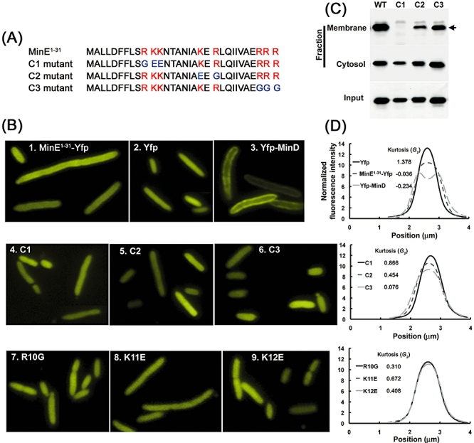
Membrane association of various mutants of MinE1–31-Yfp in Δmin cells. A. MinE1–31 contains three clusters of basic residues (red). This region of MinE1–31 was engineered to disrupt the positively charged residues in these clusters. The substituted residues are coloured in blue. B. Localization of wild-type (1) and mutant MinE1–31-Yfp (4–9) as well as untagged Yfp (2) and Yfp-MinD (3) in Δmin cells. C. Western blot analyses on fractionated cells expressing the mutant forms of MinE1–31-Yfp. The membrane fraction was estimated to be 30-fold more concentrated than the cytosolic fraction in this experiment to facilitate comparisons between different MinE1–31-Yfp bound to the membrane. The arrow indicates MinE1–31-Yfp. D. Analysis of fluorescence distribution across the cell width and G2 of MinE1–31-Yfp, Yfp and Yfp-MinD (top), mutant MinE1–31-Yfp containing C1, C2 or C3 mutations (middle) and mutant MinE1–31-Yfp containing R10G, K11E or K12E mutations (bottom).
