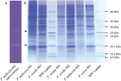
Fig. 5.
Identification on 8–18% density gradient SDS gel of stylar proteins associated with RNase activity in accessions of F. nubicola and F. viridis. (a) Part of a gel showing the track of the F. nubicola 601 native protein sample stained for RNase activity. (b) Remaining section of the gel showing concentrated samples stained with Colloidal Coomassie Blue, and Sigma low molecular weight ladder. In (a), the gel has been photo-reduced by 20% to compensate for its expansion after destaining relative to the gel in (b). The bands that correspond to RNase activity at 21 kDa and/or 30 kDa, present in the accessions of SI F. nubicola 601 and 603 and in F. viridis 901, 902, and 903 but absent from SC F. vesca 801, are marked by arrows. The bands for submission for MS–MS sequencing were excised from a similar gel initially stained for RNase activity, and then destained and restained with Colloidal Coomassie Blue.

