Fig. 2.
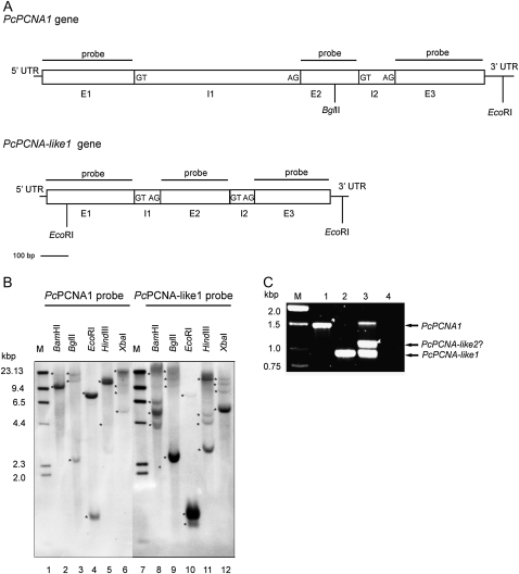
Southern blot and PCR analysis of P. coccineus genomic DNA. (A) Structure of PcPCNA1 and PcPCNA-like1 genes. The nucleotide sequences were analysed using WebGene software. Exons (E), introns (I), and 5′-UTR and 3′-UTR regions are marked. The border sequences of introns termini are placed in the boxes. The positions of internal sites recognized by restriction enzymes used in the Southern blotting analysis are marked. (B) Southern blotting results. 30 μg of the genomic DNA isolated from P. coccineus seedlings were digested with BamHI (lanes 2 and 8), BglII (lanes 3 and 9), EcoRI (lanes 4 and 10), HindIII (lanes 5 and 11) or XbaI (lanes 6 and 12), separated in 0.8% agarose gel and subjected to Southern blot procedure with the PcPCNA1 (lanes 2–6) or PcPCNA-like1 (lanes 8–12) probe. Lanes 1 and 7: DNA molecular weight marker II Digoxigenin-labelled (Roche). Stars indicate position of DNA fragments detected with PcPCNA probes. (C) PCR results. PCR was performed using degenerate primers and gDNA isolated from P. coccineus seedlings (lane 3), or plasmid pTZ57R\T DNA containing genomic sequence of the PcPCNA1 gene (lane 1) or of the PcPCNA-like1 gene (lane 2). In negative control, DNA template was omitted (lane 4). Lane M: DNA size standards (1 kb DNA ladder).

