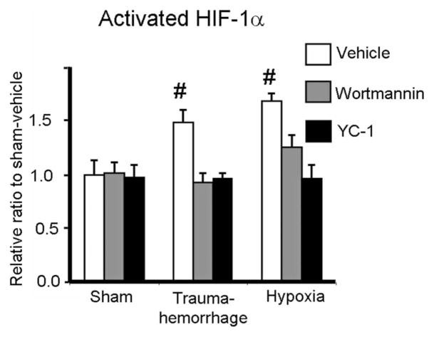
FIGURE 5.
Expression of activated HIF-1α in Kupffer cells following trauma-hemorrhage or hypoxia. Male C3H/HeN mice were subjected to sham treatment, trauma-hemorrhage or hypoxia as described in Materials and Methods. Each group of animals was given Wortmannin (1 mg/Kg body weight, BW), YC-1 (5 mg/Kg BW) or vehicle at maximum bleedout time or 30 minutes after induction of hypoxia. Upon harvesting, Kupffer cells were isolated, nuclear proteins were extracted, and HIF-1α levels were determined as described in Materials and Methods. Data are mean ± SE; n = 6 animals/group. #P < 0.05 compared with all the other groups.

