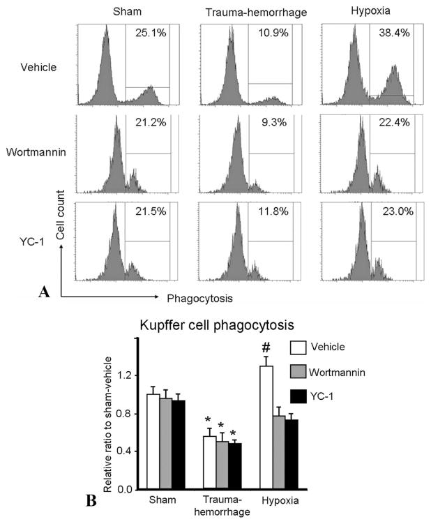
FIGURE 7.
Kupffer cell phagocytic capacities following trauma-hemorrhage or hypoxia. Male C3H/HeN mice were subjected to sham treatment, trauma-hemorrhage or hypoxia as described in Materials and Methods. Each group of animals was given Wortmannin (1 mg/Kg body weight [BW]), YC-1 (5 mg/Kg BW) or vehicle at maximum bleedout time or 30 minutes after induction of hypoxia. Kupffer cells were isolated and cultured overnight. Following overnight incubation, the cells were cultured with bioparticles for 1 hour and phagocytosis was determined as described in Materials and Methods. A, The marked numbers in the gated region of representative histograms indicate the percentage of Kupffer cells that had ingested bioparticles. B, Data are shown as mean ± SE; n = 6 animals/ group. *P < 0.05 compared with sham and hypoxia groups; #P < 0.05 compared with all the other groups.

