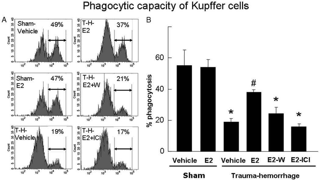
FIGURE 4.
Phagocytic capacity of Kupffer cells. Mice were subjected to sham operation or trauma-hemorrhage (T-H) and were treated with vehicle, estrogen (E2), estrogen plus Wortmannin (E2-W), or estrogen plus ICI 182,780 (E2-ICI) immediately before resuscitation. E. coli bioparticle conjugates for the phagocytosis assay were injected into each group immediately before resuscitation. Livers were harvested aseptically 2 h after resuscitation, and Kupffer cells were isolated. The Kupffer cells were surface stained with an allophycocyanin-Alexa Fluor 750-conjugated CD11b Ab as described in Materials and Methods and were analyzed by flow cytometry. The percentage of Kupffer cells that ingested bioparticles is shown by representative histograms (A) and as mean ± SE (B) for n = 6 animals/group. *, p < 0.05 compared with both sham groups; #, p < 0.05 compared with the other three trauma-hemorrhage groups.

