Figure 8.
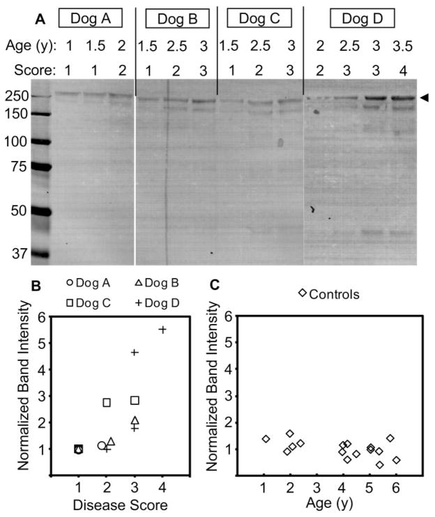
Increased ANGPTL7 protein concentration in canine AH correlated with disease progression in the beagle POAG model. Equal volumes (20 μL) of AH humor sampled from POAG beagle dogs at 6-month intervals was separated by nonreducing SDS-PAGE, followed by anti-ANGPTL7 Western blot analysis (A). At each AH sampling, the dogs were examined and scored for severity of glaucoma (1, very mild; 2, mild; 3, mild-moderate; and 4, moderate). Leftmost lane: molecular weight in kilodaltons. The band intensities were quantified and normalized to the band intensity of the earliest sample for each dog (B). The top band (A, arrowhead) was chosen for quantification. The age-dependent increase in ANGPTL7 was determined by anti-ANGPTL7 Western blot analysis of equal volumes (20 μL) of AH obtained from healthy laboratory-quality mongrel dogs aged 1 to 6 years and band intensities normalized to the average band intensity for the gel (C).

