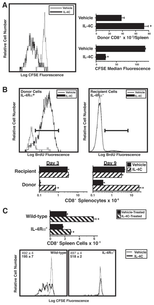
Figure 3. IL-4 acts directly on CD8+ T cells to induce proliferation and accumulation.
A. BALB/c IL-4Rα-deficient mice were injected i.v. with 1.6 × 10⁶ CFSE-labeled purified wild-type BALB/c splenic CD8+ cells, then injected i.p. every other day with vehicle or IL-4C (10 μg IL-4/60 μg anti-IL-4). Spleen cells obtained from recipient mice 3 d after cell transfer were stained for CD8, CD4 and IL-4Rα and analyzed for number and CFSE fluorescence of CD8+IL-4Rα+ cells. N = 4; * signifies p <.05 compared to cells from vehicle-treated mice.
B. BALB/c IL-4Rα-deficient mice were injected i.v. with 4 × 10⁶ purified wild-type BALB/c splenic CD8+ cells on d 0, i.v. with anti-CD4 mAb on d 0 and 6; i.p. every other day with vehicle or IL-4C (10/60); and twice i.p. 1 d prior to sacrifice with BrdU. Spleen cells from recipient mice sacrificed 3 or 9 d after cell transfer were stained for IL-4Rα, CD8, and BrdU and analyzed for number and BrdU staining of CD8+IL-4Rα+ (donor) and CD8+IL-4Rα− (host) cells. Histograms are of cells from mice sacrificed 3 d after cell transfer. N = 3–4; * signifies p <.05 compared to cells from vehicle-treated mice.
C. Wild-type BALB/c mice were injected i.v. on d 0 with 4 × 10⁷ CFSE-labeled wild-type or IL-4Rα-deficient splenic cells and injected i.p. every other day with vehicle or IL-4C (5/30). Spleen cells from mice sacrificed 3 d after cell transfer were stained for CD8 and IL-4Rα and analyzed for CFSE staining and number of donor CD8+IL-4Rα+ and IL-4Rα− cells. N = 4–5; * signifies p <.05 compared to cells from vehicle-treated mice.

