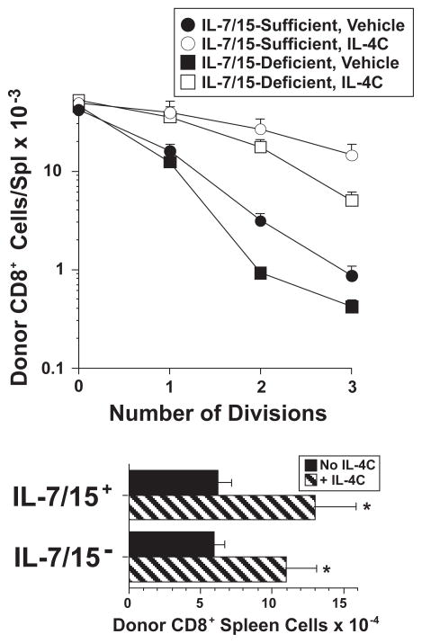
Figure 8. IL-4-induced CD8+ T cell proliferation is IL-7- and IL-15-independent.
C57BL/6 CD8+ spleen cells were purified and labeled with CFSE. 1.75 × 10⁶ of these cells were transferred into either C57BL/6 wild-type mice that had been injected 24 hr earlier with 3 mg of control mAb or C57BL/6 IL-15-deficient mice that had been injected 24 hr earlier with 3 mg of anti-IL-7 mAb. Recipient mice were injected on the day of cell transfer and 2 d later with vehicle or IL-4C (5/30) and also received a second dose of control mAb or anti-IL-7 mAb 2 d after cell transfer. Spleen cells from recipients sacrificed 3 d after cell transfer were stained for CD4 and CD8 and analyzed to determine number of CD8+CFSE+ cells and CFSE fluorescence intensity, which was then used to determine number of cell divisions. N = 3–4; * indicated p <.05 compared to CD8+ T cells from the same mouse strain that received the same treatment by received no IL-4C.

