
Figure 1.
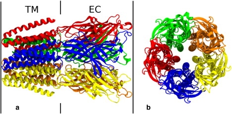
Side (a) and top (b) views of the crystal structure of GLIC2. Protein subunits are individually colored, and the transmembrane (TM) and extracellular (EC) domains are noted.
Official websites use .gov
A
.gov website belongs to an official
government organization in the United States.
Secure .gov websites use HTTPS
A lock (
) or https:// means you've safely
connected to the .gov website. Share sensitive
information only on official, secure websites.
Side (a) and top (b) views of the crystal structure of GLIC2. Protein subunits are individually colored, and the transmembrane (TM) and extracellular (EC) domains are noted.