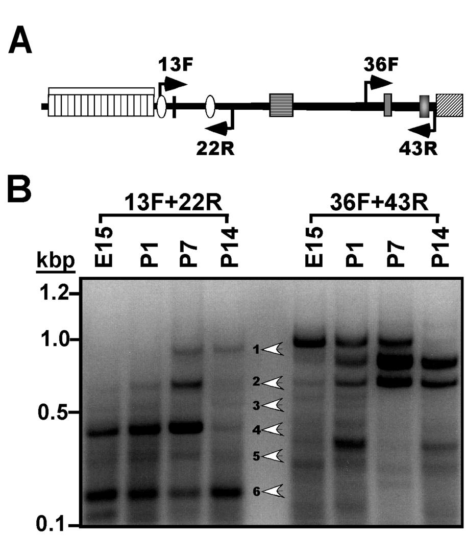
Figure 2. Developmental regulation of the expression of densin 5’ and 3’mRNA splice variants.
A. Schematic domain diagram of densin protein. Labeled arrows indicate approximate positions of primers used in this figure. See Fig. 1A for details of domain structure. B. Analysis of mRNA splicing in the 5’ and 3’ coding regions of the densin mRNA during development. Total RNA isolated from rat brains of the indicated ages (in days; E, embryonic; P, postnatal) was analyzed by RT-PCR using the indicated primers. An ethidium bromide-stained agarose gel is shown with white arrowheads indicating the 6 major products generated using the 13F+22R primer pair.

