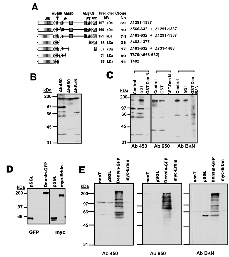
Figure 4. Expression of densin variants in adult brain.
A. Cartoon depicting domain structures of predicted densin spice variants and the locations of epitopes used to generate antibodies. The protein variant at the top is the major adult form (Apperson et al. 1996), with conserved domains and motifs annotated as in Fig. 1A. Structures below were predicted from mRNA variants summarized in Fig. 3C with the corresponding clone number listed afterwards. B. Initial testing of densin antibodies. Rat forebrain whole lysates were immunoblotted using the three antibodies (indicated in Panel A). C. Characterization of antibody specificity for endogenous densin. Antibodies were pre-incubated for 1.5 hours at room temperature with GST, GST-DenN or GST-BΔN (25 µg each in 1 ml), as indicated, and then used to immunoblot adult rat forebrain whole lysates. D. Expression of densin and erbin in transfected HEK293 cells. Myc-Erbin, densin(Δ1291–1337)-GFP, and a control myc/GFP-tagged densin protein (pSGL) were expressed in HEK293 cells and cell lysates were probed for myc or GFP. Using pSGL as a control, amounts of myc-erbin and densin-GFP lysates loaded to the gels were adjusted to contain approximately equal amounts of erbin and densin. E. Characterization of densin antibody specificity in heterologous system. myc-Erbin, densin-GFP and pSGL were expressed in HEK293 cells: nonT, non transfected cells. Gels loaded with similar amounts of erbin and densin lysates (see panel D) were immunoblotted using densin antibodies.

